About us
We provide specialist equipment and expert scientists to help you carry out analytical research.
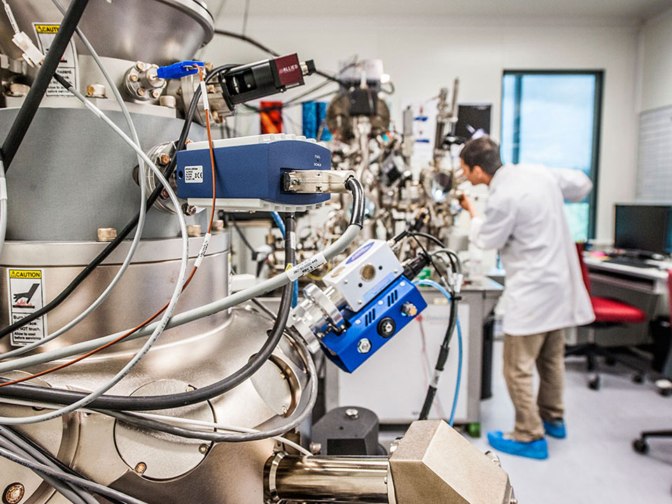
Our state-of-the-art equipment and expert technologists offer transdisciplinary analytical support, including sample preparation, data collection and interpretation of results.
The Central Analytical Research Facility (CARF) is located across QUT campuses, with the main node situated in the Science and Engineering Centre at our Gardens Point campus.
Services and equipment
We offer services to QUT staff and students, external researchers and commercial organisations, ranging from basic technical support to commercial research.
Specialist staff are available to carry out analyses, or we can train you to use the instruments to do your own work.
Prices for services and equipment use are current as at time of publishing, and are subject to change.
Contact carf@qut.edu.au to discuss your project.
Capabilities
We support scientific analytical research, teaching and learning to enable real-world results.
Cell analysis
The Cell Analysis Facility offers state-of-the art imaging and cell analysis instrumentation to facilitate imaging and analysing the molecular and structural organisation of cells, tissue and bioengineered materials.
The facility is run by qualified operators and provides comprehensive training in all aspects of microscopy and cell analysis from experimental design, sample preparation, instrument operation and data analysis.
Technologies and equipment offered
- Confocal laser scanning microscopy
- Wide-field fluorescence microscopy
- Live-Cell Imaging
- Digital deconvolution
- Transmitted-light imaging (phase, DIC)
- Specialized microscopy (FRAP, FRET)
- Scanning electron microscopy
- Flow cytometry
- Digital Spatial Profiling – GeoMx Digital Spatial Profiler – nanostring
- nCounter Analysis System for Multiplex Analysis up to 800 Targets - nCounter Prep station and Digital Analyser
Services offered
The Cell Analysis Facility provides expertise and technical resources to assist researchers in designing experimental plans, acquiring appropriate data, and interpreting data. Training is provided for staff and students to independently conduct their cell analysis research. For the infrequent, untrained user, our facility provides a completely assisted technical support service.
Contact us
For more information on what we offer and how to access our expertise please email cellanalysisfacility@qut.edu.au
Microscopes
The Cell Analysis Facility provides access to a wide range of microscope platforms including both routine and advanced optical and electron microscopes. The technology capabilities include scanning electron microscopy, bright field microscopy (phase contrast and DIC), multi-channel fluorescence microscopy, confocal laser scanning microscopy, and is fully equipped for live-cell imaging.
| Instrument | Capabilities and features |
|---|---|
| Leica TCS SP5 Confocal Microscope |
|
| Leica AF6000LX Widefield Microscope |
|
| Zeiss AxioImager Z2 Widefield Microscope |
|
| Holomonitor M4 Live Cell Imaging System |
|
| Nikon Eclipse TiS Inverted Microscope with Fluorescence |
|
| Nikon Eclipse Ts2 Routine Inverted Microscope |
|
| Hitachi TM3000 ESEM |
|
Flow cytometry
The flow cytometry resources provide multi-parameter flow cytometric analysis for the evaluation of cell proliferation and development, apoptosis, presence of internal and external cell markers and proteins of interest, cell cycle analysis and much more.
| Instrument | Capabilities and features |
|---|---|
| BD FACSCelesta Flow Cytometer |
|
| MACSQuant Flow Cytometer |
|
| BD FACSMelody Fluorescence Activated Cell Sorter |
|
| FlowJo |
|
Our experienced staff can provide training, advice and guidance on the experimental process and the analysis of results, to ensure optimal outcomes.
Spatial genomics
The Nanostring GeoMx DSP and nCounter Flex System are available through the CARF Cell Analysis Facility.
| Instrument | Capabilities and features |
|---|---|
| Nanostring nCounter Flex System |
|
| Nanostring GeoMx Digital Spatial Profiler | Digital spatial profiling (DSP) allows you to combine high-plex gene expression profiling with the spatial resolution of immunohistochemistry. This technology is useful for examining cellular interactions, tissue heterogeneity, pathogenicity and response to therapy. The GeoMx DSP workflow starts with staining of the tissue section with up to four fluorescent markers to visualize the tissue morphology and detection probes coupled to photocleavable target specific oligonucleotide tags. The tissue section is imaged and regions of interest (ROI) are selected for expression profiling, based on the morphology of the tissue. The ROIs are sequentially exposed to UV light, releasing the oligonucleotide tags, which are quantitated on the Nanostring nCounter instrument or through next generation sequencing. The counts of each target are mapped back to the tissue location, revealing the spatial expression patterns of the tissue. Supported Assays:
The protein assays and Immune pathways RNA panel use nCounter to quantitate the oligonucleotide tags whereas the larger CTA and WTA panels use next generation sequencing as read-out method. Sample requirements:
Recommended section thickness is 5um. Since DSP experiments are complex, we require an initial consultation with you so that we may assist you with planning and design of your experiment. During this initial project discussion, we will gain an understanding of your project goals, sample type, desired analyte (protein/RNA), timeline and estimated cost. The major determinants of overall project cost will be the number of desired slides to be processed, region of interest (ROI) number and size, and nCounter/sequencing requirements. We are committed to working with your to execute y our spatial profiling experiments efficiently and cost-effectively. Please contact us at spatialgenomics@qut.edu.au to discuss your project requirements. |
Our experienced staff can provide training, advice and guidance on the experimental process and the analysis of results, to ensure optimal outcomes.
Booking instructions
Contact Christina Theodoropoulos via email at cellanalysisfacility@qut.edu.au to discuss how the Cell Analysis Facility can assist in your research, and to make a booking.
External users
Cell analysis facilities are available by arrangement to staff and postgraduate students from any university and other external users. Users external to QUT can access the facility from Monday to Friday, 9am-5pm. Contact us at cellanalysisfacility@qut.edu.au to discuss your requirements and to request a quote.
Our Cell Analysis Facility is available to QUT staff and students and external clients at competitive rates.
Elements and isotopes
Overview
Our researchers can help analyse your materials and samples using the cutting-edge technologies in our elements and isotopes laboratory.
Our laboratory houses an array of top-end instruments for chemically analysing a wide range of gas, liquid and solid samples, down to parts per trillion in some cases.
Most samples need to be crushed, powdered, extracted or diluted while avoiding contamination into a form that can be analysed by the instrument. We can assist with preparing samples as required.
Details
Our state-of-the-art equipment allows for a range of analyses and discovery.
Carbon, nitrogen and sulfur (CNS) analysis
| Instrument | Capabilities and features |
|---|---|
| LECO CNS928 Series Macro Determinator | Analysis of carbon, nitrogen and sulfur in macro samples (up to 3g for nitrogen; up to 1.5g for carbon/nitrogen; up to 0.3g for carbon/nitrogen/sulfur). |
| FlashSMART CHNS or O | Analysis of hydrogen, carbon, nitrogen, oxygen and sulfur in micro samples (maximum size 2mg). |
| Shimadzu TOC-V (with TNM attachment) Total Organic Carbon Analyser | Measurement of total carbon (TC), inorganic carbon (IC), total organic carbon (TOC) and total nitrogen (TN) in liquid samples, such as river, lake, ocean, wastewater and industrial samples (max concentration ~100ppm). |
Chromatography
| Instrument | Capabilities and features |
|---|---|
| Agilent 7890A Gas Chromatograph | Analysis of methane, carbon dioxide and nitrous oxide |
| Shimadzu Nexus GC-2030 | Analysis of greenhouse gas and other permanent gases |
| Dionex Integrion Ion Chromatograph | Anion analysis (F-, Cl-, NO3-, SO42-, Br-) of aqueous samples, and solid samples (F-, Cl). Analysis methods can be made to order. |
Electrochemistry
| Instrument | Capabilities and features |
|---|---|
| TPS pH, conductivity and dissolved oxygen meter and ion probes | pH, conductivity, dissolved oxygen and specific ion measurement for aqueous samples. |
| Mettler Toledo T50 Titrator-Rondolino | Automated titration with nine-place sample changer for alkalinity, acid base titration, etc. |
Mass spectrometry
| Instrument | Capabilities and features |
|---|---|
| Sercon 20-22 Isotope Ratio Mass Spectrometer | Precise measurements of 12C, 13C, 14N, 15N, 16O, 17O, 18O isotopes in gas, liquid or solid samples. |
| Agilent 8800 Inductively Coupled Plasma Mass Spectrometer (ICP-MS) | Triple quadrupole mass spectrometer capable of trace metal analysis down to parts per trillion. |
| Agilent 8900 Laser Ablation Inductively Coupled Plasma Mass Spectrometer (LA-ICP-MS) | Determination of trace element concentrations, down to parts per billion, in a wide range of solid samples. |
| Syft Technologies VOICE200ultra Selected Ion Flow Tube Mass Spectrometer (SIFT-MS) | High sensitivity, real-time high-throughput quantitative gas analysis. Simultaneous analysis of chemically diverse VOCs and inorganics to pptv level. |
| Shimadzu TD-GCMS Thermal Desorption Gas Chromatograph Mass spectrometer | Analysis of VOCs collected in thermal desorption tubes. Shimadzu TD-30 series. |
Spectroscopy
| Instrument | Capabilities and features |
|---|---|
| Thermo Fisher Gallery Discrete Analyser | Automated wet chemistry analyser with extensive applications: water, wastewater, soil, plant tissue, fertiliser, etc. |
| Agilent Cary 60 UV-VI Spectrophotometer | Spectrometer for ultraviolet-visible analysis. Software allows for a wide range of applications. |
| Perkin Elmer Optima 8300 DV and Agilent 5800 Inductively Coupled Plasma Optical Emission Spectrometer (ICP-OES) | Elemental analysis down to parts per million. Dual view instrument for TDS up to 20% and dynamic linear range. |
| Los Gatos Research (LGR) Liquid Water Isotope Analyser | Measures δ18O and δ2H water in liquid. |
Sample preparation
| Instrument | Capabilities and features |
|---|---|
| Freeze Dryer | Drying/water removal from solid samples including sediments, plant material and soils. |
| Bench top preparation | Bench area for sample and reagent preparation. Glassware, ovens, dessicators, sinks and gases. |
| Dionex ASE 350 Accelerated Solvent Extractor (ASE) | Extraction of solid and semi-solid samples using common solvents at elevated temperatures (40-200°C) and pressures. |
| Classie TheOX electronic fusion instrument | Preparation of inorganic samples (fusion solutions and fused disks) for X-ray fluorescence (XRF). |
| Milestone Ultrawave Single Reaction Chamber (SRC) | SRC microwave digestion system, up to 199 bars of pressure and 350°C. |
| Clean room | Trace metal analysis preparation. |
| Analab CleanAcids and Savillex DST-1000 Acid Purification System | Sub-boiling stills for purification of acids and reagents such as HNO3, HCI, HF and other solvents. |
| Balance room | Various analytical balances with precision to 0.1 mg and 0.01 mg, as well as an anti-static kit. |
Services and equipment hire
Our equipment is available to staff, students, external researchers and commercial organisations at a competitive price. Our laboratory provides both self-service and full-service options.
Conditions
- A minimum number of samples applies depending on the instrument or analysis. Extra charges may apply or waiting time may be longer to make a batch.
- For non-conventional samples requiring non-routine analysis, method development or additional advice on analytical setup and data processing, an additional charge will apply.
- Charges will apply for additional sample preparation required before analysis (eg. ICPMS acid digestion, LECO dry and grind, IC extraction).
Indicative pricing for external researchers
| Equipment, service or analysis | With technician assistance | Unassisted |
|---|---|---|
| Laser ablation ICP-MS | ||
| ICP-MS equipment use (half-day, 4 hours) | $1,140 | $960 |
| ICP-MS equipment use (full-day, 8 hours) | $1,700 | $1,360 |
| ICP analysis (per sample) | ||
| ICPOES - Major and trace element | $32 | N/A |
| ICPMS - Trace and ultra trace elements | $55 | N/A |
| Acid digestions/extractions for ICP analysis (per sample) | ||
| Room temperature extraction/dilution | $12 | N/A |
| Hotblock extraction/digestion | $21 | N/A |
| Hot plate digestion | $75 | N/A |
| Microwave digestion | $120 | N/A |
| Elemental analysis (per sample) | ||
| Elemental analysis: O (mg-sized samples) | $45 | $30 |
| Elemental analysis: CHNS (mg-sized samples) | $45 | $30 |
| Bulk elemental analysis: CNS (g-sized samples) | $20 | $15 |
| TOC-TN (liquid) | $20 | $15 |
| Sample preparation (per sample) | ||
| Dionex ASE (solvent extraction) | $40 | $20 |
| Ashing | $20 | N/A |
| Sample Grinding and drying (~20g) | $25 | N/A |
| Fine milling for CHNS or O (< 1g) | $12 | N/A |
| Freeze drying (fit inside 50ml vial) | $20 | N/A |
| Nutrient analysis (per sample) | ||
| Gallery Discrete Analyser (NO3- and NH4+) | $13 | $8 |
| Anions by IC (per sample) | ||
| Dionex 2100: F-, Cl-, NO3-, SO42-, Br- | $25 | $20 |
| Integrin IC with Combustion for solids F-, Cl-, SO42- | $50 | N/A |
| Gas analysis (per sample) | ||
| CO2, CH4, N2O by GC | $6 | $5 |
| H2, O2, CO, CO2, N2, N2O2, NO by GC | $12 | $10 |
| SIFT-MS | ||
| VOICE200 Ultra (half day, 4 hours) | $530 | N/A |
| VOICE200 Ultra (full day, 8 hours) | $930 | N/A |
| Stable Isotopes Analysis (per sample) | ||
| IRMS: N2O, N2, CO2 gas by Cryo-IRMS | $10 | $9 |
| IRMS: C and N in solids by EA-IRMS | $20 | $15 |
| 2H and 18O by LWIA | $17 | $12 |
Indicative pricing for commercial organisations
| Equipment, service or analysis | With technician assistance |
|---|---|
| Laser ablation ICP-MS | |
| ICP-MS equipment usage (half-day, 4 hours) | $2,760 |
| ICP-MS equipment usage (full-day, 8 hours) | $4,100 |
| SIFT-MS | |
| Batch set-up fee | $160 per batch |
| Known analytes (up to 10 compounds) | $110 per sample |
| Unknown analytes (up to 10 compounds) | $230 per sample |
| With operator (half day, 4 hours) | $1,300 |
| With operator (full day, 8 hours) | $2,300 |
| ICP analysis (per sample) | |
| ICPOES - major and trace elements | $55 per sample |
| ICPMS - trace and ultra trace elements | $90 per sample |
| Acid digestions/extractions for ICP analysis (per sample) | |
| Room Temperature extraction/dilution | $25 per sample |
| Hot block extraction/digestion | $40 per sample |
| Hot plate digestion | $115 per sample |
| Microwave digestion | $185 per sample |
| Elemental analysis | |
| Elemental analysis: O (mg-sized samples) | $95 per sample |
| Elemental analysis: CHNS (mg-sized samples) | $95 per sample |
| Bulk elemental analysis: CNS (g-sized samples) | $40 per sample |
| TOC-TN (liquid) | $40 per sample |
| Sample preparation (per sample) | |
| Dionex ASE (solvent extraction) | $85 per sample |
| Ashing | $45 |
| Sample Grinding and drying (~50g) | $60 |
| Fine milling for CHNS or O (< 1g) | $30 |
| Freeze drying (fit inside 50ml vial) | $30 |
| Nutrient analysis (per sample) | |
| Gallery Discrete Analyser (NO3 and NH4) | $30 per sample |
| Anion by IC (per sample) | |
| Dionex 2100: F-, Cl-, NO3-, SO42-, Br- | $45 per sample |
| Integrion IC with Combustion for solids F-, Cl-, SO42- | $120 per sample |
| Stable isotopes analysis | |
| IRMS: N2O, N2, CO2 gas by Cryo-IRMS | $30 per sample |
| IRMS: C and N in solids by EA-IRMS | $35 per sample |
| 2H and 18O by LWIA | $35 per sample |
| Gas analysis (per sample) | |
| N2O, CH4, CO2 gas by GC | $15 per sample |
| H2, O2, CO, CO2, N2, N2O, NO by GC | $20 per sample |
Sample submission forms
For assistance with preparing, labelling and submitting samples, please contact us.
Genomics
Overview
We are equipped with the latest genomic technology to offer a broad range of services from sample extraction, preparation and analysis. Our experienced staff operate from both NATA -accreditation and PC2 laboratories, enabling us to accommodate a variety of samples, both clinical and research. We can provide support for all aspects of the genomics sample preparation and analysis workflow, including DNA/RNA extraction, library preparation, quality control, manual and automated sample processing, Next-Generation-Sequencing, Microarrays and bioinformatics analysis.
Our equipment is available to staff, students and external clients with both self-service and full-service options. Our experienced staff can provide training, advice and guidance on the experimental design process and the analysis of results, so that you achieve optimal outcomes.
Services
We routinely run a wide variety of services, that are continuously being upgraded as newer technologies emerge and listed below are the more commonly run services. We are also happy to work with researchers on protocol development for new methods or trialling unusual samples that may have not been tested with existing technologies.
Prices are largely dependent on sample number and will fluctuate accordingly. We are happy to help you plan your experiment so you can aim for sample numbers that give you the best value for money, whilst delivering statistically relevant results.
If you have any further enquiries in relation to the services we provide or need additional information, email carfgenomics@qut.edu.au.
| Service | per sample/per project ** | |
|---|---|---|
| Ideal sample number | AUD/sample | |
| DNA extraction - blood/saliva* | 24 | $33 |
| DNA extraction - tissue* | Please enquire | |
| DNA extraction - FFPE scrolls* | Please enquire | |
| DNA/RNA co-extraction - FFPE scrolls* | Please enquire | |
| DNA extraction (non-standard) | Prices vary depending on sample type | |
| RNA extraction (non-standard) | Prices vary depending on sample type | |
| FFPE extraction (non-standard) | Prices vary depending on sample type | |
| Whole genome sequencing | 5-60 | **$6,976-$32,784 |
| RNA sequencing | 8-192 | **$4,721-$68,801 |
| DNA sequencing | Prices vary depending on sample type | |
| Exome sequencing | 32-570 | **$14,059-$179,114 |
| Clinical Exome sequencing* | 24 | Please enquire |
| Targeted and panel sequencing | Prices vary depending on sample type | |
| Pan-Cancer panel sequencing | 24 | Please enquire |
| TSO500 HT DNA/RNA* | 24 | Please enquire |
| Hi-C sequencing | Prices vary depending on sample type | |
| Single cell sequencing | Prices vary depending on sample type | |
| Isoform sequencing | 1-4 | **$5,664-$7,752 |
| Nanopore sequencing | 1 (Multiplex) -24 | **$12-$4,964 |
| PacBio sequencing | 1 (10-18kb) - 96 (16S) | **$3,493-$4,128 |
| Microbiome sequencing | Prices vary depending on sample type | |
| Metagenomics | Prices vary depending on sample type | |
| Epigenetics | Prices vary depending on sample type | |
| Nanostring sequencing | Prices vary depending on sample type | |
| ELISA assays | Prices vary depending on sample type | |
| ChIP sequencing | Prices vary depending on sample type | |
| SNP microarray (global screening array) | 364 | **$91 |
| SNP microarray (bisulfite modification and methylation EPIC array) | 192 | **$511 |
| Bioinformatics | Arrange a consultation as early as possible for details |
* Optional NATA-accredited services
Technology and equipment
The equipment at our facilities has a range of applications and we are happy to discuss your needs to find what works best for you. Certain equipment is available for use by researchers with training available.
To find out more or discuss what you would like to do, email carfgenomics@qut.edu.au.
Quality control
| Instrument | Capabilities and features |
|---|---|
| Agilent Fragment Analyzer 5200 | Uses automated parallel capillary electrophoresis to provide reliable and accurate quality control for DNA and RNA samples. Sample types include genomic DNA, NGS libraries, small RNA, cell free DNA, large DNA fragments and total RNA quality assessment. |
| Agilent Femto pulse | Powerful and automated pulsed-field capillary electrophoresis system. Easily achieve 10 times higher sensitivity for smears and up to 100 times for fragments. A meticulously designed optical detection platform and pulsed-field power enable unprecedented sensitivity, detecting nucleic acids into the lower femtogram range. |
Tapestation 4200 | This system is an established automated electrophoresis tool for DNA and RNA sample quality control. The flexible platform allows QC of a single sample, through to a full 96-well plate while maintaining cost-efficiency. |
| Qubit 4.0 | This Fluorometer is the next generation of the popular benchtop fluorometer designed to accurately measure DNA, RNA, and protein quantity. The easy-to-use touch screen menus make it easy to select and run the assays you need, with results displayed in just a few seconds. |
Qubit Flex | A benchtop fluorometer designed for highly accurate quantification of DNA, RNA, microRNA, and protein. With the Qubit Flex Fluorometer you have the flexibility to directly measure fluorescence of up to eight samples simultaneously, which reduces assay variability. |
| Varioskan LUX microplate reader | Designed for bioscience researchers with a wide variety of needs, this microplate reader comes equipped with a flexible range of measurement technologies including Absorbance, Fluorescence Intensity, Luminescence, AlphaScreen, and Time-Resolved Fluorescence. The Varioskan LUX multimode reader has received HTRF™ certification from Cisbio Bioassays and DLReady™ validation from Promega Corporation. |
Nanodrop | DNA, RNA and protein quantitation with only 1–2 µL of sample in seconds. Frequently used alongside Qubit to determine purity of samples. |
| Corbett Rotor Gene qPCR | Instrument for quantitative real-time amplification that’s also capable of end-point analysis, nucleic acid concentration measurement and now HRM (high resolution melt). |
Trinean Dropsense | A compact, benchtop spectrophotometer for plate-based high-speed quantification of DNA, RNA or protein samples. |
Preparation
| Instrument | Capabilities and features |
|---|---|
| Nadia Instrument | An automated, microfluidic droplet-based platform for single cell research that encapsulates up to 8 samples, in parallel, in under 20 mins. Over 50,000 single cells can be captured per cartridge in a run. |
| Nadia Innovate | Enables the development of user-defined single cell protocols and applications. Newly developed protocols can be transferred to the Nadia Instrument for high throughput parallel operation. |
| QIAsymphony | A fully automated extraction system that can accommodate up to 96 specimens in the one run. This bead-based technology enables purification of DNA, RNA, and bacterial and viral nucleic acids from a wide range of starting materials. |
| Covaris E220 | A plate-based sonicator for reliable and reproducible fragmentation of samples in preparation for Next-Generation Sequencing library preparation. |
| Covaris M220 | The M220 Focused-ultrasonicator offers the “Scientist’s Standard” in a compact, easy-to-use system making it the ideal DNA shearing solution for MiSeq and PGM users. AFA® technology in the M220 eliminates operator-induced variations, improves recoveries, increases efficiency, and provides standardized results. |
| Tecan liquid handling | Innovative liquid handling technology that is flexible and reliable for high through-put SNP microarray processes. |
| Janus liquid handling | A flexible automated liquid handling robot to meet specific application needs, including high-throughput nucleic acid normalisation, NGS sample preparation and sample pooling. |
| Eppendorf epMotion 507t | Unique mixing performance with excellent temperature control to guarantee complete, dependable, and reproducible assay results. |
Sequencing/analysis
| Instrument | Capabilities and features |
|---|---|
| Pacbio Sequel | Revolutionary sequencing technology combines completeness of long reads with the accuracy of short reads to provide a comprehensive view of genomes, transcriptomes and epigenomes. |
| Nanopore MinION, MK1C, Flongle and GridION | A range of portable, self-contained sensing technology that uses nanopores embedded in high-tech electronics, to perform precise molecular analysis. Offers real-time data delivery of short to ultra-long fragments of native DNA or RNA. |
| Illumnia MiSeq | Facilitates 0.3Gb to 15Gb of output with 1-25 million sequencing reads and 50 to 600 bp read lengths. Ideal for targeted re-sequencing, metagenomics, small genome sequencing and expression profiling sequencing projects. |
| Illumina NovaSeq 6000 | Illumina’s most powerful sequencing system, designed for scalable throughput by offering output up to 6 Tb and 20 billion reads in dual flow cell mode. Flexible workflow with streamlined operations increasing efficiency at cost effective rates for HTP sampling. |
BioRad Droplet Digital PCR | ddPCR technology enables high-throughput digital PCR in a manner that uses lower sample and reagent volumes while maintaining the sensitivity and precision that are the hallmarks of digital PCR. ddPCR technology provides an absolute count of target DNA copies per input sample without the need for running standard curves, making this technique ideal for measurements of target DNA, viral load analysis, and microbial quantification. |
| Nanostring nCounter | Provides a simple and cost-effective solution for multiplex analysis of up to 800 RNA, DNA, or protein targets. Accelerate your research with just 15 minutes total hands-on time without amplification, cDNA conversion, or library prep and generate publication ready figures in ~24 hours. |
| Nanostring GeoMx digital spatial profiler | Combines the best of spatial and molecular profiling technologies by generating digital whole transcriptomes and profiling data for 100s of validated Protein analytes from up to 12 tissue slides per day. This unique combination of high-plex and high-throughput spatial profiling enables researchers to rapidly and quantitatively assess the biological implications of the heterogeneity within tissue samples. |
| Illumina iScan | High-precision scanner that supports an extensive range of array applications. This system can scan thousands of array samples per day, without sacrificing data quality or reproducibility. It supports our expansive portfolio of genetic analysis assays, from high-throughput genotyping to DNA methylation analysis. |
Using our services and equipment
We can provide support for all aspects of the genomics sample preparation and analysis workflow, including: DNA/RNA extraction library preparation quality control manual and automated sample processing bioinformatics analysis.
Our equipment is available to staff, students and external clients with both self-service and full-service options. Our experienced staff can provide training, advice and guidance on the experimental design process and the analysis of results, so that you achieve optimal outcomes.
If you need equipment not currently available in the CARF genomics laboratory, it is likely we can arrange access to this, either through our partner institutions or preferred service providers.
Histology
The Histology lab is a complete service laboratory located at our Kelvin Grove campus.
Our laboratory can process, embed, section, stain and image fresh or fixed tissue, and specialises in expert, reproducible, high throughput histological services. We offer:
- routine paraffin, cryo and more specialised resin histology services
- soft and hard tissue capacity, specialising in bone tissue histology
- rapid decalcification of mineralised samples
- client designed tissue micro array blocks
- automated immunostaining (enzyme labels, fluorescence labels, multiplexing, in situ hybridisation, NanoString protocols)
- *specialised imaging capability including brightfield and fluorescence modalities for stereomicroscopy,and light microscopy, and infrared (FT-IR) spectroscopy
- high throughput slide scanning.
Sample preparation
We work with human, animal and plant samples often with implanted medical and regenerative devices. To ensure optimal sample preparation, we can:
- rapidly decalcify hard or mineralised tissues before processing for routine paraffin histology and/or immunohistochemistry options
- embed mineralised samples in resin and section (thin or ground) to better preserve tissue morphology (ideal for bone and metallic/ceramic implants common in dentistry and orthopaedics)
- rapidly freeze and cryo section samples with labile antigens or processing-sensitive components prior to staining.
Accessing services and equipment
Our histology laboratory is available to staff and students and external clients at a competitive price. It provides both self-service and full-service tissue processing, sectioning, histochemical and immunohistochemical staining and automated, high throughput slide scanning.
Our experienced staff can provide training, advice and guidance on the experimental process and the analysis of results, so that you achieve optimal outcomes.
Booking instructions
To arrange a consultation, email histology@qut.edu.au
Conditions
- Work will not commence until a quote has been agreed upon.
- Machine use will be charged as 'per use' for any period less than an hour and in full 1-hour increments thereafter.
- Paraffin sections will be placed onto slides with 2 sections per slide, sample size permitting.
- If embedding is being undertaken, the client must be present to advise on the orientation of the sample.
- Automated staining may be set up for specific stains.
- A minimum of 10 slides is required for all paraffin sectioning and staining requests.
Indicative pricing for external researchers
Machine use
| Machine | Per hour |
|---|---|
| Exakt saw | $20 |
| Exakt grinder | $20 |
| TegraPol grinder/polisher | $20 |
| Resin microtome | $20 |
Histology services
Prices include labour unless stated otherwise.
| Service | Labour per hour | Per sample | Per slide |
|---|---|---|---|
| Tissue processing | Not applicable | $4 | Not applicable |
| Paraffin embedding | $95 | $6 | Not applicable |
| Paraffin sectioning | $95 | Not applicable | $7.50 |
| Resin embedding - 25mL mould | $95 | $75 | Not applicable |
| Resin embedding - 100mL mould | $95 | $300 | Not applicable |
| Resin ground section - large | $95 | Not applicable | $40 |
| Resin ground section - small | $95 | Not applicable | $25 |
| Automatic H&E staining and coverslipping | Not applicable | Not applicable | $6 |
| Automatic counterstain (Hx after IHC) and coverslipping | Not applicable | Not applicable | $3 |
| Other simple stains and coverslipping - Safranin O, Oil Red O, Sirius Red, Toluidine Blue, von Kossa, Trichrome, Methyl Green | Not applicable | Not applicable | $8 - $10 |
| Slide scanning (20 slides minimum) | Not applicable | Not applicable | $10 |
| Training | $95 | Not applicable | Not applicable |
Equipment
Our state-of-the-art equipment allows for:
- paraffin histology
- resin histology
- cryo histology
- high throughput processing and slide scanning
- advanced light microscopy
- image analysis
- customised tissue micro array blocks
Magnetic resonance spectroscopy
Overview
Our nuclear magnetic resonance and electron paramagnetic resonance spectrometers allow us to investigate the structure, dynamics and interactions of materials at the molecular scale.
The Magnetic Resonance lab hosts several Nuclear Magnetic Resonance (NMR) spectrometers and an Electron Paramagnetic Resonance (EPR) spectrometer.
Equipment
The laboratory specialises in two different modalities of magnetic resonance spectroscopy:
- Nuclear Magnetic Resonance (NMR) spectroscopy for the study of a range of nuclei (typically 1H, 13C, 19F, 31P) in solution-state samples
- Electron Paramagnetic Resonance (EPR) spectroscopy for the study of molecular species which contain one or more unpaired electrons, including stable free radicals, antioxidants, transition metal ions and reactive intermediates.
Nuclear Magnetic Resonance (NMR) spectrometers
| Instrument | Capabilities and features |
|---|---|
| Bruker Avance IIIHD 600 MHz | High-resolution NMR spectrometer typically configured for automated 1D and 2D spectroscopy of solution state samples under ambient conditions. Probe: 5mm broadband BBO probe (1H and 107Ag to 19F) with 2H lock channel and Z gradient. Temperature range -150 to +150°C SampleJet sample changer operating under IconNMR automation. BCU-I cooling unit for temperature control. |
| Bruker Avance IIIHD 400 MHz | High-resolution NMR spectrometer with; QNP and BBIBBO probes for 1D and 2D spectroscopy of solution state samples under ambient or variable-temperature conditions; High-Resolution Magic Angle Spinning (HR-MAS) probe for spectroscopy of gel-state samples (e.g. swollen polymers, swollen beads, tissue samples).
Liquid nitrogen dewar and boil-off heater for low-temperature measurements. |
| Bruker Avance Neo 400 MHz | High-resolution NMR spectrometer with; TBO probe for 1D and 2D spectroscopy of solution state samples under ambient or variable-temperature conditions; High-Resolution CP Magic Angle Spinning (CP-MAS) probe for spectroscopy of solid-state samples.
SampleCase sample changer operating under IconNMR automation. BCU-II cooling unit for temperature control. |
Electron Paramagnetic Resonance (EPR) spectrometer
| Instrument | Capabilities and features |
|---|---|
| Magnettech MiniScope MS400 | Benchtop EPR spectrometer with variety of sample tube holders, from capillary up to 5mm tubes. Variable temperature measurements -170 to +200°C. Access port for in-situ sample irradiation. |
Using our services and equipment
Our equipment and services are available to QUT staff and students, external researchers and commercial clients.
The Magnetic Resonance facility typically operates under a self-service (unassisted) model for trained users, with some full-service (with technical assistance) options as listed below. Our experienced staff can provide training, advice and guidance on the experimental process and the analysis of results, so that you achieve optimal outcomes.
Due to the complex nature of most Magnetic Resonance experiments, we will work with you to assess the feasibility of the analysis, timeframes and costs for your project.
Indicative pricing for assisted use
Prices are indicative and vary depending on whether work is for external researchers or commercial organisations, and who carries out the work. We will provide a costing proposal for your approval before any work starts. Bookings are limited to 2 hours during the day (8am - 5pm).
| Equipment, service or analysis | External researchers | Commercial organisations |
|---|---|---|
| NMR or EPR spectroscopy (per hour) | $200 | $495 |
| NMR or EPR instrument training (per hour) | $110 | $275 |
| Assistance with sample preparation, method development, data analysis, report preparation (per hour) | $110 | $275 |
Microscopy
Overview
Our microscopy laboratory has a suite of routine and advanced light microscopes and electron microscopes that enable users to conduct innovative research. We also offer geological sample preparation, and nanoscale imaging including ion beam microscopy.
Equipment
Our state-of-the-art equipment allows for optical microscopy, electron microscopy and specimen preparation.
Optical microscopy
Our optical microscopy instruments include stereo, petrographic, metallographic, biological and confocal microscopes. The laboratory can carry out light microscopy, scanning probe microscopy and thin film characterisation.
Light microscopy
| Instrument | Capabilities and features |
|---|---|
| Leica M125 Zoom Stereo Microscope | Image stack collection and extended focus reconstruction. LED ringlight, gooseneck lights and motorised z-stand. Also has transmitted brightfield, darkfield, and oblique illumination. |
| Nikon Eclipse LV100ND Petrographic Microscope | Polarised light optics in both transmission and reflection, rotating stage and motorised z-drive for 3D and extended depth of focus imaging. Compensator and full and quarter wave plates available for additional analysis. |
| Nikon Eclipse Ni | Upright microscope with transmitted brightfield and DIC. Motorised nosepiece and condenser for ease of use. |
| Leica DMi8A Inverted Microscope | Inverted materials microscope for metallography in brightfield and polarised light including scanning stage and manual focus drive. |
| Nikon A1R Confocal Microscope | Confocal imaging optimised for fixed biological samples. Resonant scanner. 405, 488 561 and 64038 nm lasers. |
| Nikon Eclipse Ti Inverted Microscope | Imaging of fluorescent or transparent samples. Equipped with fluorescent filters for DAPI, FITC, TRITC, and Cy5 and similar fluorescent labels. Also equipped with transmitted brightfield, DIC, and polarisating optics. Motorised stage with stage inserts for imaging large areas on single slides, or multi-well plates. |
Electron microscopy
Our electron microscopy instruments include an electron probe microanalyser (FE-EPMA), focused ion beam (FIB) microscopes, scanning electron microscopes (SEMs) and transmission electron microscopes (TEMs).
Capabilities
We can:
- embed, section, and stain fixed biological tissues or samples (SEM, TEM)
- mount and polish materials (eg. minerals, glasses, metal alloys) in thin section, epoxy resin or conductive resin (SEM, TEM, EPMA)
- perform microstructural and compositional analysis (TEM, SEM) (eg. electron diffraction, EDX on SEM and TEM)
- produce high resolution TEM images (lattice structure information)
- undertake tomography and Z-contrast imaging
- perform nanostructuring (FIB) and HRTEM lamella preparation, targeted cross-sectioning, tomography, including running simulations (SRIM, FIB/SEM)
- perform electron back-scattered diffraction (EBSD) to investigate structure and orientation of polished crystalline materials
- use wavelength dispersive x-ray spectroscopy to generate high precision standards-based quantitative elemental analysis of solid materials (EPMA)
- integrate panchromatic and hyperspectral cathodoluminescence imaging (UV- vis-near IR) into EPMA
- carry out liberated mineral analysis (LMA)
- carry out electron and ion (He, Ne) beam lithography for nanostructuring.
Electron probe microanalyser (EPMA)
| Instrument | Capabilities and features |
|---|---|
| JEOL JXA 8530F Hyperprobe | Field emission electron microprobe for high-precision, standards-based quantitative elemental analysis (Be to U) and x-ray mapping using Probe for EPMA and Probe Image software. Electron gun operates up to 30 kV and several mA beam current, with detection limits routinely ≤100 ppm and reaching <10 ppm. Twelve analyzing crystals are distributed among five wavelength dispersive spectrometers (WDS) including a large crystal "L" type and two high count rate "H" types. Detector array features two sealed Xe and three gas-flow proportional counters. Additional capabilities for imaging and qualitative analysis include a Thermo Noran System 7 EDS system, JEOL panchromatic cathodoluminescence (CL) detector, and both standard and high-resolution xCLent spectroscopic CL detectors. Anti-contamination cold finger is available. |
Scanning electron microscopes (SEMs)
| Instrument | Capabilities and features |
|---|---|
| Zeiss Sigma VP Field Emission Scanning Electron Microscope | Zeiss Gemini column gives excellent imaging performance across the beam energy range for imaging delicate structures and surface features. Variable Pressure (VP) operation for insulating samples. Oxford XMax 50 Silicon Drift (SDD) EDS detector for rapid X-ray mapping and accurate analysis. |
| JEOL 7001F Scanning Electron Microscope | A very stable instrument capable of delivering very high beam currents in a small spot size for X-ray and electron backscatter diffraction (EBSD) analysis. Automated feature detection and analysis software. Field emission gun, Oxford XMax 80 X-ray detector, Inca gunshot residue (GSR), Oxford EBSD system. |
| Hitachi SU7000 Scanning Electron Microscope | A high-resolution imaging SEM with stage biasing for low-voltage imaging of delicate structures and surface features and variable pressure mode for insulating samples. Equipped with dual in-column detectors, a chamber SE detector, and a 5-segment retractable BSE detector, offering multi-signal and simultaneous display. Fitted with Oxford Ultim Max 100 EDS detector for rapid X-ray mapping and accurate microanalysis. Aztec GSR gunshot residue software package is included as well. |
| TESCAN Mira3 Scanning Electron Microscope | Stage biasing for low-voltage imaging of delicate structures and surface features. Variable Pressure (VP) mode for insulating samples available with a cold stage. Fitted with a 60 mm² Thermo Ultra Dry EDS Detector. The microscope has an Electron Beam Lithography package for nanostructuring. |
| TESCAN TIMA Field Emission Scanning Electron Microscope | An automated mineral analysis system that fully integrates backscattered electron (BSE) and cathodoluminescence (CL) imaging with energy dispersive X-ray (EDX) spectrometry for fast quantitative analysis of rock samples and natural resource materials such as ores, tailings and smelter products. |
| Phenom XL G2 Desktop Scanning Electron Microscope | Contains a large sample holder with motorised stage navigation, low/medium/high vacuum modes, light optical and electron optical (BSE and SE) modes. The Phenom includes an EDS detector with integrated software. The resolution of this microscope is excellent due to the thermionic (CeB6) source (<10 nm). |
Focused ion beam instruments (FIB and HIM)
| Instrument | Capabilities and features |
|---|---|
| Zeiss ORION NanoFab Helium Ion Microscope (HIM) | High-resolution scanning ion microscope that employs high energy ion beam (helium or neon) for sub-nanometer imaging and nanofabrication. HIM is equipped with electron flood gun for charge compensation and imaging of nonconductive materials. The use of dual-ion beams (such as helium and neon) allows for a wide range of applications, including nanoscale ion implantation and defect engineering. |
| TESCAN S8000x Xe Plasma FIB-SEM | FIB-SEM instrument that combines Xenon plasma FIB with field-free UHR FE-SEM, for characterization of materials in large (up to 0.5 mm in width) FIB cross sections, multi-modal FIB-SEM tomography for 3D reconstruction (including 3D EDS and EBSD analysis) and fabricating micro- and nanostructures, using the implantation-free Xe ion beam (e.g. TEM lamellas and micropillars for mechanical testing). |
Transmission electron microscopes (TEMs)
| Instrument | Capabilities and features |
|---|---|
| JEOL 2100 200 kV Transmission Electron Microscope | High-resolution imaging capabilities for a range of materials and nanotechnology applications. High-sensitivity silicon drift X-ray detector (Oxford XMax) for compositional analysis. Ultra-high-resolution pole piece, TVIPS Fx416 4K CCD camera, LaB6 gun. |
| JEOL 1400 120 kV Transmission Electron Microscope | High-contrast optics, optimised for imaging of biological samples, and high tilt range for acquisition of tomographic datasets. TVIPS F4216 2K CCD camera, tomography holder, cryo holder, tungsten gun. |
| JEOL ARM200F "NeoARM" | Aberration Corrected Transmission Electron Microscope. Capable of sub-angstrom imaging and sub-nanometre compositional analysis of a wide range of samples. High resolution pole piece, capable of supporting high tilt range for tomography experiments. Equipped with Gatan OneView camera and Cold Field Emission Gun. |
Sample preparation
| Instrument | Capabilities and features |
|---|---|
| Gatan Cryoplunge 3 | Cryofixation of biological samples for transmission electron microscopy. |
| Pelco Biowave Pro Microwave Tissue Processor | Microwave accelerated processing of tissue samples for scanning electron microscopy and transmission electron microscopy. |
| Leica EM UC6 Ultramicrotome | Sample preparation of embedded biological samples for transmission electron microscope characterisation. |
| Leica EM UC7 Ultramicrotome | Sample preparation of embedded biological samples for transmission electron microscope characterisation. |
| Leica EM TXP | Target preparation device for milling, sawing, grinding and polishing samples. |
| Gatan Model 691 Precision Ion Polishing System (PIPS) | Precise ion milling of samples for transmission electron microscopy. |
| Gatan Model 682 Precision Etching and Coating Systems (PECS) | Ion milling of light microscope and scanning electron microscope samples up to 25 mm diameter mounted blocks. High quality sputtered coatings of chromium and carbon. |
| Leica EM-SCD005 Sputter Coater | Gold sputter coating. |
| Leica EM ACE600 High Vacuum Coaters | Two ACE600 coaters - one for platinum sputter coating and another with dual heads for carbon thread coating or iridium sputter coating. |
| Safematic CCU-010 HV High Vacuum coater | Carbon thread coating. |
| Tousimis Autosamdri-815 Critical Point Drier (CPD) | Automated CPD for dehydration of samples for electron microscopy. |
| Evactron 25 Decontaminator with Softclean Chamber | Gentle removal of hydrocarbon contamination from samples and components by oxygen free-radical stream. Can also be used to glow discharge sample surfaces for a hydrophilic finish to improve fluid mounting. |
| E.A. Fischione Model 2000 Specimen Preparation System | Precision grinding of samples for transmission electron microscopy. |
| E.A. Fischione Model 330 Ultrasonic Disk Cutter | Cutting undamaged 3mm disk samples for transmission electron microscopy. |
| Fischione Model 110Twin Jet Electropolisher | Thinning 3mm metal samples for transmission electron microscope (TEM) imaging. |
Using our services and equipment
Our microscopy equipment is available to staff, students, external researchers and commercial organisations at a competitive price.The laboratory provides both self-service and full-service options, with comprehensive instrument training available for internal and external academic researchers as well as commercial clients. Our experienced staff can provide advice and guidance on the experimental procedures and design and the analysis of results in order to achieve optimal outcomes.
Indicative pricing
Prices are indicative and vary depending on whether work is for external researchers or commercial organisations, and who carries out the work. We will provide a costing proposal for your approval before any work starts.
Indicative pricing for external researchers
| Equipment, service or analysis | With technician assistance | Unassisted |
|---|---|---|
| SEM microscopy (per hour) | ||
| JEOL 7001F | $165 | $75 |
| Hitachi SU7000 | $165 | $75 |
| Zeiss Sigma | $165 | $75 |
| TESCAN Mira 3 | $165 | $75 |
| TESCAN TIMA | $165 | $75 |
| Phenom XL G2 | $165 | $75 |
| Electron probe microanalysis (per hour) | ||
| JEOL 8530F | $165 | $75 |
| TEM microscopy (per hour) | ||
| JEOL 1400 | $165 | $75 |
| JEOL 2100 | $165 | $75 |
| JEOL ARM200F "NEOARM" | $165 | $75 |
| Interpretation and reporting | $100 | N/A |
| Ion microscopy (per hour) | ||
| Helium Ion Microscopy | $200 | $100 |
| Xe Plasma FIB | $200 | $100 |
| Interpretation and reporting | $100 | N/A |
| Other microscopy (per hour) | ||
| Light microscopes | $135 | $50 |
| Nikon A1R Confocal | $165 | $75 |
| Other services (per hour) | ||
| Microscopy preparation | $100 | N/A |
Indicative pricing for commercial organisations
| Equipment, service or analysis | With technician assistance | Unassisted |
|---|---|---|
| SEM microscopy (per hour) | ||
| JEOL 7001F | $415 | $285 |
| Hitachi SU7000 | $415 | $285 |
| Zeiss Sigma | $415 | $285 |
| TESCAN Mira 3 | $415 | $285 |
| TESCAN TIMA | $415 | $285 |
| Phenom XL G2 | $415 | $285 |
| Electron probe microanalysis (per hour) | ||
| JEOL 8530F | $415 | N/A |
| TEM microscopy (per hour) | ||
| JEOL 1400 | $415 | N/A |
| JEOL 2100 | $415 | N/A |
| JEOL ARM200F "NEOARM" | $415 | N/A |
| Ion microscopy (per hour) | ||
| Helium Ion Microscopy | $550 | N/A |
| Xe Plasma FIB | $550 | N/A |
| Interpretation and reporting | $200 | -N/A |
| Other microscopy (per hour) | ||
| Light microscopes | $230 | $105 |
| Nikon A1R Confocal | $295 | $180 |
| Other services (per hour) | ||
| Microscopy preparation | $130 | N/A |
| Materials preparation | $130 | N/A |
| Consulting and report writing | $260 | N/A |
Molecular mass spectrometry
Overview
Our laboratory houses cutting-edge mass spectrometry instrumentation interfaced with liquid chromatography and gas chromatography capabilities. This equipment enables selective identification and reproducible quantification of trace-level molecules in complex samples.
Equipment
Our state-of-the-art equipment allows for:
- liquid chromatography mass spectrometry (LC-MS)
- gas chromatography mass spectrometry (GC-MS)
- sample preparation.
Liquid chromatography mass spectrometry (LC-MS)
| Instrument | Capabilities and features |
|---|---|
| Shimadzu Triple Quadrupole 8050 LC-MS/MS |
High-sensitivity and ultra-fast polarity switching platform with ESI and APCI ionisation capabilities. Chromatography: Nexera UPLC. Application: Targeted quantitation of trace-level compounds (eg. nucleotides, pharmaceuticals, metabolites and lipids). |
| SCIEX Triple Quadrupole Trap 6500 LC-MS/MS platform |
High-sensitivity platform for targeted, quantitative MRM-based assays. Also features differential ion mobility (DMS) capabilities for enhanced separation/structural elucidation of isobars and epimers. Chromatography: Prominence HPLC. Application: Shotgun lipidomics, quantitation of peptides. |
| Thermo Fisher LTQ Orbitrap Elite |
High-resolution MS/MS platform equipped with an automated nanospray robot. Chromatography: Dionex UPLC. Application: Untargeted discovery of synthetic molecules and endogenous metabolites. |
| Thermo Fisher LTQ-XL |
Linear ion trap MS/MS platform with photodissociation capabilities. Chromatography: Dionex UPLC. Application: Low resolution identification of synthetic molecules. |
| Waters Synapt G2-Si |
Ion-mobility enabled quadrupole time of flight platform, equipped with LC-ESI or Desorption Electrospray Ionisation (DESI) ion sources. Chromatography: Waters Acquity i-Class UPLC. Application: Mass spectrometry imaging via DESI, ion-mobility MS. |
| Waters Select Series Cyclic IMS |
Ultra-high resolution ion mobility platform for unparalleled resolution of isobars and isomers. Features ESI and DESI ion sources. Chromatography: Waters Acquity Premier UPLC. Application: Ion mobility separation of isomeric molecules of synthetic or biological origin. |
Gas chromatography mass spectrometry (GC-MS)
| Instrument | Capabilities and features |
|---|---|
| Shimadzu Triple Quadrupole 8040 GC-MS/MS |
Capable of high sensitivity, highly reproducible, MRM-based quantitation of trace-level compounds. Equipped with high-efficiency ion source.
Auto-sampler: liquid injection, SPME, headspace, automated derivatization. Application: Metabolite profiling with online derivatization. |
| Shimadzu Triple Quadrupole 8050 GC-MS/MS |
Capable of quantitative analyses of ultra-trace level compounds, down to the femtogram level. Equipped with high efficiency detector.
Auto-sampler: liquid injection, SPME, headspace, automated derivatization. Application: Quantitative analysis of environmental pollutants, volatiles, residual solvents and metabolites. |
| Shimadzu QP2020 with Pyrolyzer |
Multi-functional pyrolyzer with vertical micro-furnace enables analysis of almost all sample forms.
Application: Analysis of insoluble materials and compounds, trace level polymer materials without pre-treatment. |
Sample preparation
| Instrument | Capabilities and features |
|---|---|
| Benchtop preparation | Includes ultra-high purity solvents and chemicals, derivatisation reagents, shakers, incubators and temperature-controlled centrifuges. |
| Martin Christ Rotational vacuum concentrator | Drying or concentrating samples with or without heating. |
| Eppendorf Thermomixer | Programmable heating, cooling and mixing. Adapters for various tube and plate formats available. |
| Perkin Elmer JANUS G3 MDT Automated Workstation | Liquid handling system for automated sample preparation. |
Using our services and equipment
Our equipment and services are available to QUT staff and students, external researchers and commercial clients with both self-service (unassisted) and full-service (with technical assistance) options.
Due to the complex nature of most mass spectrometry experiments, please email carfproteomics@qut.edu.au to discuss your project requirements.
All prices are ex GST.
Indicative pricing
Liquid chromatography-mass spectrometry (LC-MS) and gas chromatography-mass spectrometry (GC-MS)
| Service description | External researchers/Collaborative | Commercial organisations |
|---|---|---|
| Analysis using established methods | ||
| Data acquisition, analysis and report |
$275 first sample $85 each additional sample |
$685 first sample $200 each additional sample |
| Sample preparation |
$440 first sample $30 each additional sample |
$1,100 first sample $75 each additional sample |
| Analysis requiring method development | ||
| Pilot acquisition, analysis and report | from $5,500 | from $13,750 |
| Sample preparation (pilot investigation) | from $2,200 | from $4,950 |
| Method validation | by agreement | by agreement |
Terms and conditions
- Testing Services agreement is required.
- Pricing above applies to assisted use only.
- Method validation against client requirements.
- Additional costs might include chromatography columns, analytical standards and other specialist consumables.
Hourly access fee
Liquid chromatography-mass spectrometry (LC-MS) and gas chromatography-mass spectrometry (GC-MS)
| Service description | External researchers | Commercial organisations |
|---|---|---|
| Access | $100/hour | N/A |
| Training | $210/hour | N/A |
Annual access fee (facility subscription)
Liquid chromatography-mass spectrometry (LC-MS) and gas chromatography-mass spectrometry (GC-MS)
| Service description | External researchers | Commercial organisations |
|---|---|---|
| Access and training | $6,950 | $16,950 |
Terms and conditions
- The training fee is for unassisted use, and does not cover assistance in sample preparation, method development and data interpretation. A separate arrangement must be made for these services.
- Requires CARF induction and Work in CARF agreement.
- Fee covers access to the mass spectrometry laboratory for a single user and up to 300 hours of instrument time which are to be used within 12 months from completing CARF induction.
- Includes 2 days of instrument training and ongoing consultation with CARF team during the subscription period.
- Subscriptions are paid in advance. Unused hours cannot be transferred , extended, or reimbursed.
- Users are responsible for booking instrumentation, data acquisition and interpretation, and purchasing project-specific consumables (e.g., autosampler vials, chromatography columns and analytical standards).
Physical and mechanical properties
Overview
Our laboratory analyses the physical and mechanical properties of any item. The lab allows researchers to characterise a material's:
- electromagnetic properties
- mechanical properties, including hardness and tensile strength
- surface area, porosity and particle size
- thermal properties (measuring changes up to 1600°C)
- electrochemical properties.
Equipment
Our state-of-the-art equipment allows for:
- electromagnetic characterisation
- mechanical properties characterisation
- surface area, porosity and particle analysis
- thermal analysis
- sample preparation.
Mechanical properties characterisation
| Instrument | Capabilities and features |
|---|---|
| Hysitron TI 950 TriboIndentor | Nanochemical characterisation and testing instrument, with dual-head testing capability for nano/micro scale connectivity. |
| Anton Paar MCR302 Rheometer | Flow properties of low to high visuosity materials in Temp range between 30°C to 200°C. |
| Struers Duramin-40 AC3 hardness tester | Performs Vickers and Knoop hardness tests in a force range from 10 g to 62.5 kg. |
| Tytron 250 Microforce Testing System (MTS) | Low-load tensile test machine rated up to 250 N. Capable of standard and fatigue testing and three-point flexural testing. Available load cells are 250 N and 10 N. |
Surface area, porosity and particle analysis
| Instrument | Capabilities and features |
|---|---|
| Malvern Mastersizer 3000 | Particle size measurements for suspended particles 1mm to 0.1 micron. |
| Malvern Zetasizer Nano ZS | Particle size and zeta potential for suspended particles <1000 nm. |
| Micromeritics 3-Flex Surface and Porosity Analyzer | Surface area (BET), pore size distribution (<800 angstroms) of solid materials using gas absorption/desorption. Nitrogen, argon carbon dioxide. Best for micropores. |
| Micromeritics ASAP 2020 Surface Area and Porosity Analyzer | Surface area (BET), pore size distribution (<800 angstroms) of solid materials using gas absorption/desorption. Nitrogen, argon carbon dioxide. Best for micropores. Chemisorption using hydrogen and oxygen also available. |
| Micromeritics Tristar II 3020 Surface Area and Porosity Analyzer | Surface area (BET), pore size distribution (<800 angstroms) of solid materials using nitrogen or argon. Three measurement ports. |
| Quantachrome Pentapyc 5200 He Pycnometer | True solid density with helium gas entering connected pores as small as 0.2 nm. |
Thermal analysis
| Instrument | Capabilities and features |
|---|---|
| Netzsch 402C Dilatometer | A vacuum-tight pushrod dilatometer for determining thermal length expansion in solids and powders up to 2000°C. |
| Netzsch STA 449F3 Jupiter Simultaneous Thermal Analyser | Simultaneous thermogravimetric analysis (TGA) and differential scanning calorimetry (DSC). Measurement of mass changes and thermal effects between 25°C and 1650°C. GCMS and gas phase FTIR analysis of evolved gases are available. |
| Netzsch LFA 467 Hyperflash | Thermal diffusivity and conductivity, and specific heat capacity measurements in the temperature range of room temperature to 500°C. |
| Netzsch HFM 436 Lambda Heatflow meter | For thermal conductivity measurements on insulating materials. Temperature range 30°C to 50°C. |
| Netzsch Phoenix DSC | For differential scanning calorimetry (DSC) measurement of samples between -90 °C and 400 °C. DSC during optical curing can be measured on this instrument. |
Thermoelectrics
| Instrument | Capabilities and features |
|---|---|
| Ulvac ZEM3 Seebek coefficient | Measures Seebek coefficient of thermoelectric materials. |
| Ulvac PEM2 | Measures power efficiency of thermoelectric materials. |
Sample preparation
| Instrument | Capabilities and features |
|---|---|
| Micromeritics Smart Vac Prep | Sample preparation for surface area/porosity analysis. |
| Micromeritics VacPrep 061 Degasser | Sample preparation for surface area and porosity analysis through gas absorption/desorption, with vacuum mode that prepares samples by heating and evacuation. |
| Sample splitters | Riffle splitter for representative dry sample preparation. Funnel splitter for representative sampling from samples in aqueous suspension. |
| Sieves | 1.5 mm and 2.4 mm sieves for preparation of samples for Mastersizer. |
| Tube furnace | 25mm diameter tube furnace, capable of 1200°C, controlled atmosphere. |
| Plasma cleaner | Diener plasma cleaner for cleaning contamination from sample surfaces. |
Using our services and equipment
Our equipment is available to staff, students, external researchers and commercial organisations at a competitive price. Our laboratory provides both self-service and full-service options.
Our experienced staff can provide training, advice and guidance on the experimental process and the analysis of results, so that you achieve optimal outcomes.
Indicative pricing
Prices are indicative and vary depending on whether work is for external researchers or commercial organisations, and who carries out the work. We will provide a costing proposal for your approval before any work starts.
Indicative pricing for external researchers (per hour)
CARF technician assistance for the equipment incurs an additional $80 per hour for external researchers, which is indicated in the costs below. External researchers wishing to operate the instruments unassisted must attend initial training which will incur training costs.
| Equipment, service or analysis | With technician assistance (per hour) | Unassisted (per hour) | Minimum booking period (hours) | Training cost¹ |
|---|---|---|---|---|
| Hysitron TL950 Nanoindentor | $185 | $105 | 4 | $3,360 |
| Kratos Axis Supra Photoelectron Spectrometer | $185 | $105 | 2 | $2,520 |
| Netzsch Jupiter Simultaneous Thermal Analysis | $165 | $85 | 4 | Price on request (sample dependent)2 |
| Anton Paar M302 Rheometer | $165 | $85 | 1 | $680 |
| ASAP 2020 BET | $165 | $85 | 4 | $1,025 |
| Tristar II 3020 BET | $165 | $85 | 4 | $1,025 |
| Netzsch LFA 467 Hyperflash | $165 | $85 | 4 | $680 |
| Netzsch DIL 402C dilatometer | $165 | $85 | 4 | $340 |
| Malvern Mastersizer | $125 | $45 | 1 | $790 |
| Malvern ZetaSizer (sizing) | $125 | $45 | 1 | $525 |
| Malvern ZetaSizer (zeta potential) | $125 | $45 | 1 | $790 |
| Vickers Microhardness Tester | $125 | $45 | 1 | $265 |
| Quantachrome He pycnometer | $125 | $45 | 4 | $790 |
| Netzsch HFM | $125 | N/A3 | 4 | N/A3 |
Table footnotes:
- Price total for training include CARF technician assistance and instrument time.
- Pricing depends on the properties of the sample.
- Instrument unavailable for independent operation by external users.
Indicative pricing for external researchers (per sample)
CARF technicians always operate the instruments for pricing per sample. Independent operation by external users is unavailable.
| Equipment, service or analysis | Price (per sample) |
|---|---|
| Netzsch Jupiter Simultaneous Thermal Analysis < 1000 C | $265 |
| Netzsch Jupiter Simultaneous Thermal Analysis > 1000 C | $370 |
| Malvern Mastersizer (incl SEM image, minimum 4 samples) | $75 |
| Malvern Mastersizer (no SEM image, minimum 4 samples) | $40 |
| Malvern Zetasizer (sizing, incl SEM image, minimum 4 samples) | $75 |
| Malvern ZetaSizer (sizing, no SEM image, minimum 4 samples) | $40 |
| Malvern Zetasizer (Zeta potential, minimum 4 samples) | $75 |
Indicative pricing for commercial organisations
CARF technician assistance for the equipment incurs an additional $155 per hour for commercial organisations, which is indicated in the costs below. Organisations wishing to operate the instruments unassisted must attend initial training which will incur training costs.
| Equipment, service or analysis | With technician assistance | Unassisted |
|---|---|---|
| Particle analysis (per sample) | ||
| Particle size distribution (laser diffraction) - no SEM image | $100 per sample | POA |
| Particle size distribution (laser diffraction) - including SEM image (meets ISO 13320:2009E) | $205 per sample | POA |
| Particle size distribution (nano particles) - no SEM image | $100 | POA |
| Particle size distribution (nano particles) - including SEM image (meets ISO 13320:2009E) | $205 | POA |
| Zeta potential (nano particles) | $150 | POA |
| Density determination using He pycnometry (min 4 hours) | $300 per hour | POA |
| Specific surface area and porosity (per sample) | $365 | POA |
| Particle analysis instrument hire (per hour) | ||
| Malvern Mastersizer (laser diffraction) | $300 | $115 |
| Malvern Zetasizer (DLS) | $300 | $115 |
| Quantachrome He Pyncnometer | $300 | $115 |
| Mechanical testing | ||
| Microhardess testing - Vickers | $300 | $115 |
| Nanoindentation studies (per hour, min 4 hours) | $460 | $285 |
| Tensile tester (250N) | $300 per hour | POA |
| Rheology studies | $360 | POA |
| Thermal analysis | ||
| Oxidation Induction Temperature | $270 | POA |
| Low temperature DSC: - 60 to 500 ℃. | $270 | POA |
| Simultaneous thermal analysis (DSC and TGA) up to 1000 ℃ | $525 | POA |
| Simultaneous thermal analysis (DSC and TGA) above 1000 ℃ | $830 | POA |
| Specific heat capacity | $900 | POA |
| Thermal conductivity by laser flash (includes specific heat capacity) | $1,050 | POA |
| Coefficient of thermal expansion, Tg, sintering temperature by dilatometry | $1,050 | POA |
Proteomics service
Overview
We use robotic sample preparation, data-independent acquisition enabled mass spectrometry and deep learning-based algorithms for rapid and simultaneous determination of the status of thousands of proteins in the sample to understand the molecular processes that are fundamental to life and wellbeing.
Details
Mass spectrometry
| Instrument | Capabilities and features |
|---|---|
| SCIEX TripleTOF 5600+ LC-MS/MS Mass Spectrometer | High-sensitivity, high-resolution and high mass accuracy discovery and quantitation platforms. Application to qualitative (DDA) and quantitative (DIA) analysis of peptides as surrogates of proteins. |
| SCIEX TripleTOF 6600 LC-MS/MS Mass Spectrometer |
Sample preparation
| Instrument | Capabilities and features |
|---|---|
| JANUS G3 liquid handling platform | Automated digestion and desalting of protein samples. |
Bioinformatics
| Instrument | Capabilities and features |
|---|---|
| High performance computing | Cloud-based software tools for DDA and DIA data processing. |
Using our services and equipment
Our equipment is available to staff, students, external researchers and commercial organisations at a competitive price. Our laboratory provides both self-service and full-service options. Our experienced staff can provide training, advice and guidance on the experimental process and the analysis of results, so that you achieve optimal outcomes.
Indicative pricing for external researchers (all prices exclude GST)
| Service description | Assisted1/CARF Drives | Unassisted2/User Drives |
|---|---|---|
| 50-200 samples (maximum 300 hours) | ||
| Sample preparation | $10,300 per batch | $3,000 per kit |
| Mass spectrometry | $7,300** per year | |
| Bioinformatics (DDA and DIA) |
Report for DDA included Report for DIA negotiable | Access included |
| 10-50 samples (maximum 75 hours) | ||
| Sample preparation | $5,150 per batch | $1,500 per kit |
| Mass spectrometry | $3,650** per year | |
| Bioinformatics (DDA and DIA) |
Report for DDA included Report for DIA negotiable | Access included |
| 1-10 samples (maximum 15 hours) | ||
| Sample preparation | $1,550 per batch | N/A |
| Mass spectrometry | $155** per sample | |
| Bioinformatics (DDA and DIA) |
Report for DDA included Report for DIA negotiable | Access included |
Indicative pricing for commercial organisations (all prices exclude GST)
| Service description | Assisted1/CARF Drives | Unassisted2/User Drives |
|---|---|---|
| 50-200 samples (maximum 300 hours) | ||
| Sample preparation | $13,950 per batch | $3,000 per kit |
| Mass spectrometry | $10,950 per year | |
| Bioinformatics (DDA and DIA) |
Report for DDA included Report for DIA negotiable | Access included |
| 10-50 samples (maximum 75 hours) | ||
| Sample preparation | $6,975 per batch | $1,500 per kit |
| Mass spectrometry | $5,475 per year | |
| Bioinformatics (DDA and DIA) |
Report for DDA included Report for DIA negotiable | Access included |
| 1-10 samples (maximum 15 hours) | ||
| Sample preparation | $3,550 per batch | N/A |
| Mass spectrometry | $355 per sample | |
| Bioinformatics (DDA and DIA) |
Report for DDA included Report for DIA negotiable | Access included |
** Difference between external and internal pricing covered by MOU with UQ, Griffith, USC, USQ, CQU, JCU, SCU and TRI
Terms and conditions
1 Assisted (automation service):
- covers submitting undigested samples as a single batch up to a specified number of samples or hours (whichever is reached first) at once
- includes protein extraction, digestion (FASP), desalting (StageTip) and mass spectrometry data acquisition by CARF staff
- CARF induction not required
- access to the laboratory and to the software tools not provided
- DIA (quantitative) batches include one DDA run of PBQC (pooled) sample
- DDA (qualitative) batches will be searched against sequences supplied by the user
- method development (i.e. non-typical prep, targeted acquisition) and quantitative report negotiable
- approx. turnaround time: 4-6 weeks.
2 Unassisted (instrument and cloud subscription):
- covers submitting digested and desalted samples in multiple batches up to a specified number of samples or hours (whichever is reached first) over the period of 12 months
- includes mass spectrometry data acquisition by CARF staff
- user to prepare samples and analyse data (generic protocols provided)
- subject to completion of CARF induction
- only one user obtains access to the laboratory
- access expires after 12 months from submitting of the first batch
- payment is non-refundable and non-transferrable if at least one batch has already been submitted
- includes free unlimited runs of QC-BSA and assistance with BSA data processing
- training (i.e. in sample prep, data analysis) negotiable
- approx. turnaround time: 2-3 weeks.
Sample preparation kit:
- only available in combination with one of the unassisted access options
- covers access to bench top equipment, consumables and chemicals (user to prepare all required solutions as per relevant protocols) required for protein extraction, digestion (FASP), and desalting (StageTip) over the period of 12 months
- subject to completion of additional laboratory induction
- access expires 12 months after submitting the first batch.
Sample preparation
Overview
Our geological rock preparation facility is based at the Banyo Pilot Plant and consists of a rock crushing and milling lab, and a geological thin section preparation lab run by specialist technicians.
Equipment
We have specialist equipment for preparing thin section samples and rock crushing.
Thin section sample preparation
| Instrument | Capabilities and features |
|---|---|
| Large cut-off machine with 300mm diamond blade | Can section large rocks and intricate shaped rock samples |
| Grinding and lapping machine | Accurate to 0.1 microns |
| Vacuum impregnation machine | High-powered unit for porous samples |
| Grinding and polishing machine | Can achieve a high-quality polish suitable for Scanning Electron Microscope (SEM) or microprobe |
| Diamond grinder and precision trim saw | High throughput with minimal loss or defamation of the sample |
Rock crushing
| Instrument | Capabilities and features |
|---|---|
| Jaw crusher | Able to crush samples from 100mm to 5mm |
| Disc mill | Able to mill samples from 40mm to 1mm |
| Ring mills with agate, tungsten carbide and steel milling heads | High and low speed milling for X-ray diffraction or X-ray fluorescence |
| Sieve shakers | Sizes from 4mm to 32 microns |
Services and equipment hire
We assist students, researchers and commercial organisations to prepare geological rock samples for scientific analysis. Our services are provided on a fee-for-service basis. We will discuss your requirements and timeframes and prepare a costed proposal for your approval before commencing any work.
Indicative pricing for external researchers
| Equipment, service or analysis | Cost (excluding GST) |
|---|---|
| Thin section 28 x 48mm ground and cover slipped | $55 per sample |
| Thin section 26 x 76mm ground and cover slipped | $55 per sample |
| Thin section 50 x 76mm ground and cover slipped | $110 per sample |
| Thin section 28 x 48mm polished | $70 per sample |
| Thin section 26 x 76mm polished | $70 per sample |
| Thin section 50 x 76mm polished | $165 per sample |
| Polished epoxy mounts 25mm diameter | $95 per sample |
| Polished epoxy mounts 30mm diameter | $105 per sample |
| Polished epoxy mounts 40mm diameter | $110 per sample |
| Crushing | $35 per 100g sample |
| Milling | $35 per 100g sample |
| Screening | $35 per 100g sample |
Indicative pricing for commercial organisations
| Equipment, service or analysis | Cost (excluding GST) |
|---|---|
| Thin sections | |
| Thin section 28 x 48mm ground and cover slipped | $95 per sample |
| Thin section 26 x 76mm ground and cover slipped | $95 per sample |
| Thin section 50 x 76mm ground and cover slipped | $140 per sample |
| Thin section 28 x 48mm polished | $130 per sample |
| Thin section 26 x 76mm polished | $130 per sample |
| Thin section 50 x 76mm polished | $210 per sample |
| Extra impregnation (thin sections) | $35 per sample |
| Blue dye impregnation | $35 per sample |
| Mounted samples | |
| Polished mounted samples 25mm diameter | $125 per sample |
| Polished mounted samples 30mm diameter | $145 per sample |
| Polished mounted samples 40mm diameter | $170 per sample |
| Cutting - core, slabs, rock into billets | $45 per sample |
| General sample analysis | |
| Dry preparation of water sensitive material | $40 per sample |
| Slabs cut, ground and sealed with epoxy | $60 per sample |
| Gravel, sand, particles or chips into thin sections | $40 per sample |
| Sample drying (sample received wet or damp) | $25 per 100g |
| Sample crushing (XRD) or pulverising (XRF) | $75 per 100g |
| Sample splitting (sub-sampling, gross sample mass greater than 100g) | $75 per 100g |
| Sample screening/sizing (client to specify size, if required) | $75 per 100g |
| Batch courier charge (applies to all sample crushing/splitting/screening) | $55 per sample |
Surface analysis
Overview
Our laboratory houses the only helium ion microscope (HIM) and ultra-high vacuum scanning probe microscope (UHV-SPM) in Queensland. Our helium ion microscope allows researchers to visualise the outer layers of almost any solid material. Turning up the beam current allows the etching of nanoscale patterns and structures.
Details
Our state-of-the-art equipment allows for surface analysis and scanning probe microscopy.
Surface analysis
| Instrument | Capabilities and features |
|---|---|
| Kratos Axis Supra | XPS system for automated analysis of sample surfaces, revealing elemental composition and oxidation states. The system features a monochromated Al Kα X-ray source and a helium lamp for UPS measurements on conduction bands. Samples may be sputter cleaned and depth profiled with the argon gas cluster ion source. The system also features sample heat and cool plus sensitive sample (glove box) sample transfer module. |
| Iontof M6 | Latest-generation ToF-SIMS instrument for high-sensitivity elemental and molecular mapping of sample surfaces. Bismuth analytical sources and argon gas cluster ion source for depth profiling of organic and other delicate samples. |
Scanning probe microscopy
| Instrument | Capabilities and features |
|---|---|
| Omicron Multiscan Lab Ultra-high Vacuum Variable Temperature Scanning Probe Microscope (UHV VT-STM) | An ultra-high vacuum system supporting a wide range of surface science and nanotechnology applications. AFM and STM measurements at temperatures from 25 to 600 Kelvin, X-ray photoelectron spectroscopy (XPS), k-cell and e-beam evaporators, argon sputtering, sample preparation and growth at temperatures up to 1000°C. |
| Bruker Icon Dimension | Multimodal AFM with ScanAsyst technology to automatically tune scan parameters in peak-force tapping mode. Quantitative mechanical property mapping (Peak-force QNM), and Kelvin Probe scanning. |
| Bruker Dektak | Stylus profilometer for simple 1-D and 2-D measurements of sample flatness, roughness and surface feature dimensions. |
Thin film characterisation
| Instrument | Capabilities and features |
|---|---|
| Bruker Dektak XT-A Stylus Profiler | An automated stylus profilometer capable of 0.5nm repeatability. Fitted with a 2 micron radius diamond stylus and a 50mm ceramic vacuum chuck. Software allows 3D mapping, 2D stress analysis and stitching of scans up to 150mm. |
| JA Woollam M2000 UI Spectroscopic Ellipsometer | Measures film thickness in single- and multi-layer systems, as well as refractive index and dielectric properties. Duel quartz-halogen and deuterium lamps allow spectral measurements from 245nm to 1690nm. Focusing optics for small spot analysis. |
Other techniques
| Instrument | Capabilities and features |
|---|---|
| JA Woollam M2000UI Ellipsometer | Spectroscopic ellipsometer for determination of thin film thickness in the micron to nanometer range. Can also be used to determine the film’s optical parameters. |
| QSense Quartz Crystal Microbalance | Quartz crystal microbalance system for adsorption and film property measurements from solution. Can be coupled to the ellipsometer for in-situ monitoring of optical properties. |
| Biolin Theta Flex Optical Tensiometer | High precision measurement system for determining fluid contact angles and surface free energy. Cradle system allows measurement from 0-95 degrees tilt. |
Services and equipment hire
Our equipment is available to staff, students, external researchers and commercial organisations at a competitive price. Our laboratory provides both self-service and full-service options.We provide sample preparation and support to suit your individual preference and budget.
Our experienced staff can provide training, advice and guidance on the experimental process and the analysis of results, so that you achieve optimal outcomes.
Indicative pricing for external researchers
| Equipment, service or analysis | With technician assistance (per hour) | Unassisted (per hour) |
|---|---|---|
| Atomic force microscopy (per hour) | $200 | $100 |
| XPS and UPS | $200 | $100 |
| ToF-SIMS | $200 | $100 |
| Surface profilometry | $180 | $80 |
| Drop shape analysis | $180 | $80 |
| $80Ellipsometry | $180 | $80 |
| Quartz crystal microbalance | $180 | $80 |
| Interpretation and reporting | $100 | - |
Indicative pricing for commercial organisations
| Equipment, service or analysis | With technician assistance (per hour) |
|---|---|
| Atomic force microscopy (per hour) | $560 |
| XPS and UPS | $560 |
| ToF-SIMS | $560 |
| Surface profilometry | $400 |
| Drop shape analysis | $400 |
| $80Ellipsometry | $400 |
| Quartz crystal microbalance | $400 |
| Interpretation and reporting | $200 |
Synchrotron science
Overview
The Australian Synchrotron has unique scientific analytic capabilities with applications in Earth and environmental sciences, advanced materials, chemistry and biology.
Equipment
Beamline capabilities
The Australian Synchrotron currently has several individual experimental stations, known as beamlines, each with specialist capabilities. Current beamlines include:
| Powder diffraction | Powerful X-ray diffraction used to study the crystallographic structure of material |
| Macromolecular crystallography | Single crystal X-ray diffraction to determine chemical and biological structures and analyse proteins, viruses and nucleic acids |
| X-ray fluorescence microscopy | Elemental mapping at the micron scale |
| X-ray absorption spectroscopy | Oxidation state and atomic coordination environment analysis suitable for chemistry, biology and materials science |
| Small and wide angle X-ray scattering | Offers shape and size distribution from the nano- to micro-scale |
| Soft X-ray spectroscopy | Provides surface-sensitive oxidation state and molecular arrangements |
| Infrared microspectroscopy | High-resolution infrared microscopy |
| THz/Far Infrared | High-resolution infrared spectroscopy |
Accessing the Synchrotron
QUT researchers are able to apply to access advanced instrumentation facilities including beamline capabilities at the Australian Nuclear Science and Technology Organisation (ANSTO) and Australian Synchrotron, based in south-east Melbourne. Access to the Australian Synchrotron is free of charge for users through a peer-reviewed competitive merit system. In addition, the Australian Synchrotron provides successful proposals with travel funding and accommodation for up to three researchers.
For more information and assistance with the merit access process, please contact Research Infrastructure Specialist (Synchrotron Science), Dr Michael Jones.
Vibrational spectroscopy
Overview
We have a range of Fourier-transform infrared spectroscopy (FT-IR) and Raman microscopes, spectrometers and accessories, and can offer expert service and advice.
Equipment
There are two main methods used to measure molecular vibrations:
- Infrared spectroscopy (IR or FT-IR) which involves the absorption of an infra-red photon
- Raman spectroscopy which involves the inelastic scattering of higher energy photons.
Materials with strong IR spectra tend to give weak Raman spectra and vice versa. Thus some systems are more suited to study by one technique over the other, particularly when the different sampling requirements are taken into account. Our expert staff can advise the most suitable method for your research.
Infrared spectroscopy
| Instrument | Capabilities and features |
|---|---|
| Nicolet iS50 FT-IR Spectrometer with Continuum IR microscope | In built diamond ATR. Extensive searchable IR libraries. Continuum IR microscope with computerised X,Y,Z-stage. Chemical mapping to spatial resolution of 10 microns possible. Near-, Mid- and Far-IR capability, D-TGS and MCT detectors. Multiple accessories. |
| Nicolet iS50 FT-IR Spectrometer | Emission accessory attached 100 – 1000°C. In situ chemical reaction kinetics, mineral transformations including NIR diffuse reflectance. |
| Nicolet Nexus 870 | Step-scan spectrometer. D-TGS and MCT detectors. Near-, Mid- and Far-IR capability. |
Capabilities
- IR Microscopy, confocal, high spatial resolution in transmission mode for thin films, reflectance mode for polished surfaces or coatings on reflective surfaces (gold silver, polished metal), and micro Ge-ATR mode for polished heterogeneous or small samples, down to 10 microns. Diamond anvil cell for even smaller quantities.
- Extensive searchable polymer library for polymer ID (>5700 spectra), common materials and forensics (>6500 spectra) and small molecules (>9000 spectra).
- Kinetics - monitor reactions in real time.
- IR accessories for every sampling requirement: diamond-ATR, Ge-ATR, variable angle and polarised Ge-ATR, diffuse reflectance, specular reflectance; grazing angle specular reflectance, Imaging ATR, and photoacoustic spectroscopy.
- IR chemical mapping of surfaces and films to 10 mm resolution, by peak or functional group or spectral decomposition methods.
- Emission IR. Monitor reactions 100-1000°C, complement thermal measurement (DSC/TGA) by measuring the IR spectrum as a function of temperature to monitor the chemical transformations of the solid.
Raman spectroscopy
| Instrument | Capabilities and features |
|---|---|
| Witec Confocal Raman microscope | 532 and 785 nm excitation. Rapid chemical imaging to 0.3 µm spatial resolution. Confocal (depth) to better than 3 µm. High precision piezo XYZ mapping stage 200x200x20 µm. |
| Renishaw Qontor Raman microscope | 355, 532, 633 and 785 excitation with photoluminescence capable filters. Spatial resolution <1 µm. Confocal to 5 µm. High speed encoded stage for chemical mapping. Linkham Heating (ambient to 1100°C), Heating and Cooling (77K-500K) and Oxford Microstat He (3.2K to 500K) stage. |
| Bruker FT-Raman spectrometer | 1064nm excitation. Low resolution XY view stage. |
Capabilities
- Raman spectroscopy with a range of excitations (355, 532, 633, 785 and 1064 µm) and an emphasis on microscopy for small or heterogeneous samples.
- Raman imaging (chemical mapping) of surfaces and films to <-0.5 µm spatial resolution, by peak or functional group or spectral decomposition methods.
- Mineral analysis, polymer curing, characterisation of 2D and 3D carbon (eg. graphene, diamond like films), semiconductor characterisation, inter and intramolecular interactions, and biomaterials.
Using our services and equipment
Our equipment is available to staff, students and external clients at a competitive price with both self-service and full-service options. Our experienced staff can provide training, advice and guidance on the experimental process and the analysis of results, so that you achieve optimal outcomes.
Indicative pricing for commercial organisations
| Equipment, service or analysis | With technician assistance | Unassisted |
|---|---|---|
| ATR FTIR spectra (per sample) | $160 | N/A |
X-ray analysis
Overview
Our X-ray analysis laboratory provides X-ray Powder Diffraction (XRPD), and X-ray Fluorescence (XRF) characterisations and their related sample preparation services.
We analyse synthetic materials (for example, chemicals, steel, alloys, polymers, pharmaceuticals, nanomaterials, materials for energy applications) and natural materials (for example, soils, rocks, minerals). We support QUT researchers, collaborate with external researchers, and provide analytical consultancy to commercial organisations in the real world.Equipment
X-ray diffraction (XRD)
X-ray diffraction (XRD) is a technique that uses the unique diffraction pattern 'fingerprint' from any solid crystalline material for qualitative identification and quantitative abundance determination of phases in mixtures of synthetic or natural materials. Typical XRD applications include qualitative and quantitative mineralogical characterisation of mine ores, mine site remediation and road construction materials etc. Using an internal or external standard with a phase mixture also allows the quantification of the amorphous (non-diffracting) content present in the mixture. Depending on the diffraction intensity of the target phase and the mixture, working detection limits are around 1 wt.% for powder XRD.
A range of X-ray sources, optics and hardware configurations are used to optimise diffraction pattern acquisition for various analyses (for example, powder, thin film, coatings, bulk, single crystal) and phase types.
Typical XRD applications in CARF X-ray Lab include:
- Powder diffraction (Phase ID, QPA, Lattice Parameter, Size/Strain)
- Clays
- Pharmaceuticals
- Co/Fe/Mn-rich sample measurements using soft X-rays
- capillary transmission (structure solving & refinement)
- in situ non-ambient diffraction (80K to 400K, RT to 1200℃, for Ref. & Trans. geometries)
- GID for polycrystalline thin films & depositions on low background Si wafer
- X-ray Reflectometry (for multi-layer thickness & interlayer roughness)
- High Resolution XRD, Reciprocal Space Mapping for single crystal thin films
- Wide-Range Reciprocal Space Mapping to define single crystal orientations
- Residual Stress (iso- & side- inclination), Texture (Pole Figure) for bulk materials
- Micro-Diffraction (χ-φ oscillation), Phase Mapping
- Pair Distribution Function (for amorphous & crystalline).
Equipment
| Instrument | Radiations | Capabilities and features |
|---|---|---|
| Bruker D8 Advance XRD | Cr, Co, Mo | Powder diffractometer with Dynamic Beam Optimisation (Motorised Divergence Slit, Motorised Air-Scatter Knife, Dynamic Detector Opening). Parallel beam/thin film GID. 90-position sample changer; PSD (Energy Resolution <330eV) with 0/90 ⁰ mount; Non-ambient Chamber; Chi-Phi-Z Cradle; PolyCapillary micro-diffraction; Texture; Residual Stress; Capillary spinner. |
| Rigaku SmartLab XRD | Cu, Mo | Powder diffractometer with parallel beam/thin film capability. 36-position sample changer; Mirrors for Divergent, Parallel, Focusing beams; Chi-phi cradle; 0D/1D/2D Pixel Area Detector; Capillary/foil Transmission; X-ray Reflectometry; Channel-cut monochromator Ge(220*2). |
| Rigaku SmartLab XRD | Cu, Mo | Powder diffractometer with parallel beam/thin film capability and small area micro-diffraction; Anton-Parr 1200⁰C oscillation stage with spinning capillary attachment; Reactor-X 1000⁰C Infrared heater; Oxford Cryostream system (down to 80K); LINKAM lowT chamber for reflection (down to 80K). |
X-ray fluorescence (XRF)
X-ray fluorescence (XRF) is an established X-ray technique for the quantitative chemical (elemental) characterisation of samples using the characteristic X-ray emission/s of the element/s XRF can be used to characterise crystalline and non-crystalline powders, bulk samples, and liquids, including oils etc.
Sample compositions can be measured using fused glass discs, pressed powder pellets or loose powders depending on specific requirements (qualitative or quantitative data, major, minor or trace level elemental abundances).
The XRF Application packages in CARF include:
- GEO-QUANT Advance: Turn-key solution for 21 oxides forms (Na2O, MgO, Al2O3, SiO2, P2O5, SO3, K2O, CaO, TiO2, V2O5, Cr2O3, Mn2O3, Fe2O3, NiO, CuO, ZnO, SrO, ZrO2, BaO, HfO2, PbO) common in geological samples, based on calibrations from 32x CRM, 5x QC, 3x DC
- METAL-QUANT: Turn-key solution for 23 elements (Al, Si, P, S, Ti, V, Cr, Mn, Fe, Co, Ni, Cu, Zn, As, Zr, Nb, Mo, Sn, Sb, Ta, W, Pb, Bi) common in metals & alloys, with reference to 3x QC, 11x DC
- PETRO-QUANT: Turn-key solution for 31 elements (Na, Mg, Al, Si, P, S, K, Ca, Ti, V, Cr, Mn, Fe, Co, Ni, Cu, Zn, As, Br, Zr, Mo, Ag, Cd, In, Sn, Sb, Ba, W, Tl, Pb, Bi) common in gasoline/ oil liquids, with reference to 5x QC
- QUANT-EXPRESS: Factory calibrated semi-quantitative elemental analysis (F to U) for any unknown samples with Fundamental Parameter Modelling.
Routine suites of elements can be measured and quantified, including:
- major and minor elements: Si, Ti, Al, Fe(III), Mn, Mg, Na, Ca, K, P, S(VI), plus Cl, F, Ba, Sr, Zr, Hf, Ni, Cu, Zn, and Pb (to nominal working detection limits of <<100ppm)
- 41 select trace elements from Sc to U (to nominal working detection limits of 15 to <1 ppm, element dependant).
Typical XRF applications include major and trace element analysis of rocks and soils, iron and sulphide ores, mine wastes and remediation materials, cement, pigments and other components in food packaging, trace element analysis of foodstuffs, analysis of precipitates from contaminated processing water, determination of elemental ratios in nanomaterials.
Equipment
| Instrument | Capabilities and features |
|---|---|
| Bruker S8 Tiger Series II Wavelength Dispersive X-ray Fluorescence (WD-XRF) Spectrometer | 4kW Rh tube, 7 Analysing crystals, collimators, tube filters, scintillation, gas flow detectors. 75 position auto-sample changer. Capable of analysing C to U. Routine LLD <100ppm most major elements, <1-2ppm most trace elements in rocks and soils. Automated unattended operation allowing quantitative major and trace element analysis on fused glass discs, pressed powder pellets, and qualitative elemental identification via wavelength scanning. |
Sample preparation
We have access to a wide range of sample preparation equipment, including fusion apparatus, and crushing, grinding and micronising equipment. Our qualified and experienced geo-analytical staff can also provide thin section preparation and petrologic investigation capabilities. Prior to sample preparation (for example, grinding or heating) and submission, please discuss XRD and/or XRF analysis and sample requirements with our expert laboratory staff.
Equipment
| Instrument | Capabilities and features |
|---|---|
| McCrone Micronising Mills | Reduce particle size to < 5 µm. |
| Rocklabs Swing Mill | Reduce particle size to < 75 µm. |
| Claisse Le Neo | Fused discs preparation for XRF. |
| Claisse TheOX electronic fusion instrument | Preparation of inorganic samples (fusion solutions and fused disks) for X-ray fluorescence (WD-XRF and ED-XRF). |
| Rocklabs Swing Mills and plate crusher | Rock preparation and crushing (located at Banyo Pilot Plant). |
| Retsch Mixer Mill | Cryo-mill at LN2 temperature. |
| Drying ovens | Drying of samples (40°C, 100°C). |
Using our equipment and services
Our equipment is available to researchers and external clients at a competitive price with both full-service and self-service options. Our experienced staff can provide training, advice and guidance on the experimental process and the analysis of results, so that you achieve optimal outcomes.
Indicative pricing for external researchers
| Equipment, service or analysis | With technician assistance (excluding GST) | Unassisted (excluding GST) |
|---|---|---|
| X-ray diffraction (XRD) equipment use | ||
| Bruker D8 Advance | $145 per hour | $80 per hour |
| Rigaku Smartlab Cu/Mo - Routine 382 | $145 per hour | $80 per hour |
| Rigaku Smartlab Cu/Mo - Research 383 | $155 per hour | $95 per hour |
| X-ray fluorescence (XRF) services | ||
| Major elements (fused glass disc) | $30 | $25 |
| Trace elements (pressed pellet) | $50 | $25 |
Indicative pricing for commercial organisations
| Equipment, service or analysis | With technician assistance (including GST) | Unassisted (including GST) |
|---|---|---|
| X-ray diffraction (XRD) services | ||
| Quantitative powder XRD | $340 per sample | N/A |
| Quantitative powder XRD with qualitative clays | $415 per sample | N/A |
| Stand alone qualitative clay analysis | $145 per sample | N/A |
| Optional additional clay treatments (client to specify treatment/s) | $50 per treatment | N/A |
| Quantitative fine fraction XRD analysis (suitable for bulk clays and bulk respirable silica) | $475 per sample | N/A |
| X-ray instrument hire rates | ||
| X-Ray Diffractometer - high throughput Co/Cu/Mo | $325 per hour | $200 per hour |
| X-Ray Diffractometer - Cu/Mo non-ambient | $375 per hour | $220 per hour |
| X-ray fluorescence bulk sample chemical analysis | ||
| Major elements - 21 elements reported as wt% oxides, includes LOI | $60 per sample | N/A |
| Trace elements - 40 elements reported as ppm (ug/g) | $55 per sample | N/A |
Our staff
- Position
- Professor
- Division / Faculty
- School of Earth & Atmospheric Sciences
- Research fields
- Geochemistry
- Analytical Chemistry
- Geology
- cm.allen@qut.edu.au
- Position
- Professor
- Division / Faculty
- School of Law
- Research field
- Law
- k.tranter@qut.edu.au
- Position
- Associate Professor/ARC Mid-Career Industry Fellow
- Division / Faculty
- School of Chemistry & Physics
- Research fields
- Inorganic Chemistry
- Environmental Science and Management
- Materials Engineering
- y.xi@qut.edu.au
- Position
- Principal Research Infrastructure Specialist (Electron Microscopy)
- Division / Faculty
- Research Portfolio
- Research fields
- Biochemistry and Cell Biology
- Other Physical Sciences
- jamie.riches@qut.edu.au
- Position
- Manager, Central Analytical Research Facility
- Division / Faculty
- Research Portfolio
- david.harris@qut.edu.au
- Position
- Research Fellow
- Division / Faculty
- School of Earth & Atmospheric Sciences
- john.caulfield@qut.edu.au
- Position
- Research Fellow
- Division / Faculty
- Research Portfolio
- Research fields
- Genetics
- Neurosciences
- n.maksemous@qut.edu.au

- Position
- Research Fellow (Proteomics)
- Division / Faculty
- School of Biology & Environmental Science
- Research fields
- Biochemistry and Cell Biology
- Medical Biochemistry and Metabolomics
- pawel.sadowski@qut.edu.au
- Position
- Postdoctoral Research Fellow
- Division / Faculty
- School of Mech., Medical & Process Engineering
- fernanda.zamboni@qut.edu.au
- Position
- Senior Research Officer (Electron Microprobe)
- Division / Faculty
- School of Earth & Atmospheric Sciences
- Research field
- Geology
- henrietta.cathey@qut.edu.au
- Position
- Research Officer (Genomics)
- Division / Faculty
- School of Biology & Environmental Science
- Research fields
- Genetics
- Agricultural Biotechnology
- Industrial Biotechnology
- kevin.dudley@qut.edu.au
- Position
- Genomics Project Coordinator
- Division / Faculty
- Research Portfolio
- victoria.coyne@qut.edu.au