A team of biomedical engineers from Australia and Germany studied how human and mouse cells communicate with each other and found that this humanized tissue is physiologically integrated into single functional bone tissue which retains species-specific ultrastructural differences.
- Humanized bone used to study cancer, autoimmunity, infections and graft vs host disease.
- Study found humanized tissue is physiologically integrated but retains species-specific ultrastructural differences.
QUT Distinguished Professor Dietmar W Hutmacher, from QUT/IHBI said the multiscale analysis, carried out by the team of biomedical engineers from QUT and scientists from the Department of Biomaterials at the Max Planck Institute of Colloids & Interfaces investigated the underlying design and morphology of the hybrid nature of humanized bone.
“Humanized bone is an organ that has been tissue engineered by implanting human bone progenitor cells combined with a 3D printed scaffold and bone growth factors,” Professor Hutmacher said.
“A major challenge in biomedical research and drug discovery is the lack of easy access to clinically relevant model organisms to study many medical conditions including cancer, autoimmunity, infections, and graft vs host disease.
“In particular, humanized mouse bone has been studied in primary and secondary bone cancer research to create a human-like bone microenvironment because it can interact with human cancer cells and demonstrate the cascade of events in tumour growth and progression.”
Professor Hutmacher said the research community knows that human and mouse bone architecture differs in many ways but the hybrid nature of humanized bone, while fundamental to research, had not been well-studied.
“It is particularly important to know just how much of the humanized bone is actually human because if human cancer cells interact with only the mouse component of the humanized bone it defeats the purpose of the model for progressing biomedical research.
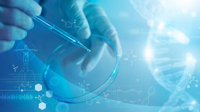
“We used cutting edge advanced-material science techniques and comprehensive immunohistochemistry to dissect species-specific differences at the cellular level to determine how much of humanized bone is ‘human’.
“We need this understanding because we know changes in bone composition and structure affect the onset of breast and prostate cancer bone metastasis and play a critical role in conditions such as osteoporosis.
“In determining the human component of humanised bone we kept in mind that both human cells and human extracellular matrix (network of proteins providing structure and biochemical support to surrounding cells) can be found in humanized bone while the vascular make-up of the bone remains mouse-derived.
 “Using advanced materials science techniques, the researchers found that humanized bone is formed by a mosaic of human and mouse collagen that is seemingly structurally integrated within the same bone organ.
“This means that both mouse and the implanted human progenitor cells contribute to tissue formation in humanized bone.
“We also investigated the extracellular matrix properties of specific human and mouse collagen regions inside the humanized bone and showed for the first time that a human-like osteocyte network is retained within the human collagen regions and is distinct from the mouse host tissue.
“Our results show the value of validating immunohistochemical analysis against a multiscale material science-based methodology to further develop and enhance human-like physiology in humanized mouse models.”
Human and mouse bone physiologically integrate in a humanized mouse model while maintaining species-specific ultrastructure was published in Science Advances.


