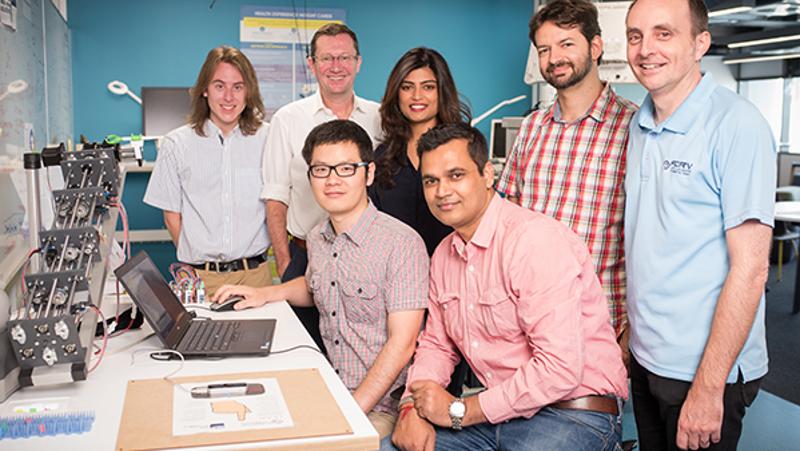
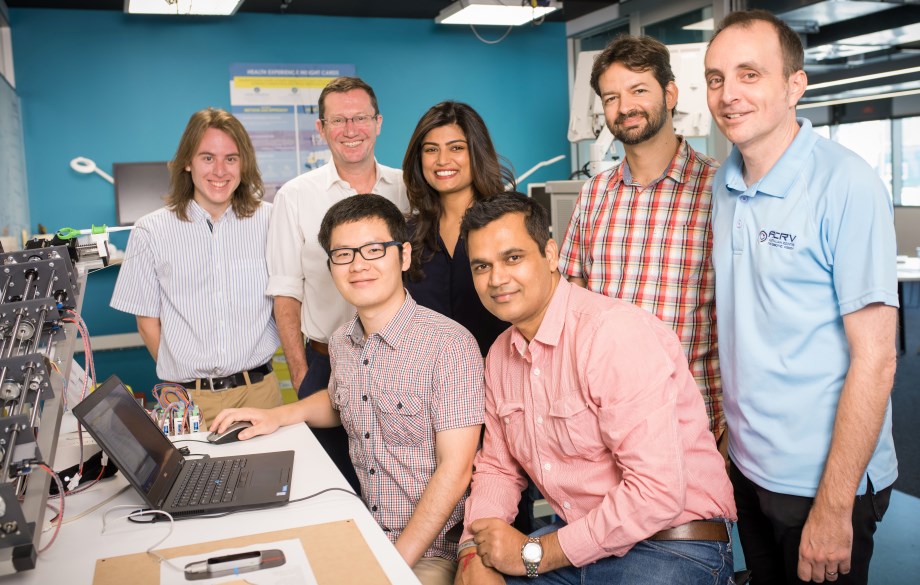
QUT is spearheading the development of a new class of medical robotics that can see soft tissue, to make keyhole surgeries simpler, safer and cheaper.
The university is leading an international collaboration that has just received a $996,000 Australia-India Strategic Research Fund grant from the Australian Government.
Project leader and renowned orthopaedic surgeon Professor Ross Crawford said the new robotic imaging system will allow surgeons for the first time to track the position of soft tissue in real time and in 3D.
“Patients recover from keyhole surgeries more quickly than open surgeries because the surgery is minimally invasive,” said Professor Crawford, from QUT’s Institute of Health and Biomedical Innovation and the Australian Centre for Robotic Vision.
“However, keyhole surgeries are very complex for surgeons to learn and perform due to the sheer difficultly in seeing into and navigating their instruments through the tiny spaces inside the body. Even experienced surgeons can cause complications to patients when performing these procedures.
“The robotic imaging system we’re building will vastly improve both visualisation and access issues, making keyhole surgery more accurate than ever before.
“We expect the rate of unintended injury during surgery will drop significantly and the training process for surgeons to be faster and safer. This is particularly important in India, where keyhole surgery is only just emerging as a mainstream medical procedure.
“But it’s the patients throughout the world who will be the biggest beneficiaries, through better outcomes and access to healthcare.”
Professor Crawford said, unlike current medical imaging tools which can only track the position of bone and medical tools, the new system will combine state-of-the-art miniaturised stereo cameras, 4D ultrasound sensing and artificial intelligence.
This will give the surgeon an accurate, real-time 3D model of the surgical site, and one that tracks the position of soft tissue like tendons and ligaments as well as bone and medical tools.
“Combining vision and ultrasound can provide us with vastly more information that we can use to identify and differentiate the objects within the surgical site,” Professor Crawford said.
“Working with a dynamic 3D model rather than a flat image on a screen is also a real game changer in terms of accuracy in keyhole surgeries – it will give the surgeon precise knowledge of how deep the objects in the surgical site really are.
“We’re building the system for knee arthroscopy surgeries first but we’re confident this is a system that can be easily adapted for other surgeries – hip, shoulder, abdominal, heart.”
QUT will partner with researchers from the Indian Institute of Technology-Madras, All India Institute of Medical Sciences-Delhi, University of Adelaide and Perfint Healthcare, in collaboration with Indian Institute of Technology-Kharagpur and Manipal University, to develop the robotic medical imaging system.
Professor Crawford said the project reflected QUT’s focus on advancing medical robotics.
Media contacts
Kate Haggman, QUT Media, 07 3138 0358, kate.haggman@qut.edu.au
After hours Rose Trapnell, QUT Media team leader, 0407 585 901, media@qut.edu.au
QUT is part of a national collaborative group of five major Australian universities that form the ATN (Australian Technology Network of Universities).


